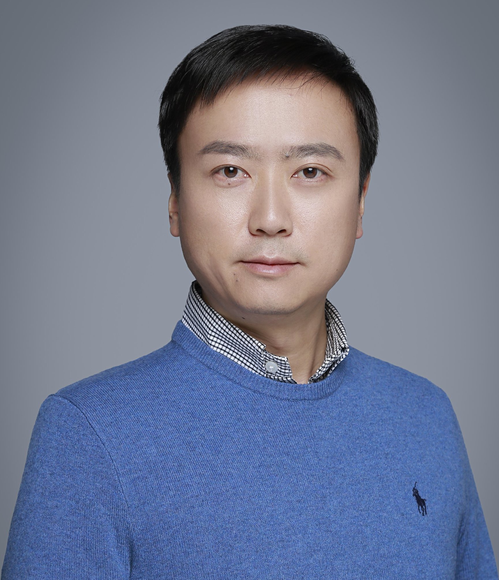
基本信息

李栋 男 博导 中国科学院生物物理研究所
电子邮件: lidong@ibp.ac.cn
通信地址: 北京市朝阳区大屯路15号
邮政编码: 100101
电子邮件: lidong@ibp.ac.cn
通信地址: 北京市朝阳区大屯路15号
邮政编码: 100101
招生信息
招生专业
071011-生物物理学
080300-光学工程
071009-细胞生物学
080300-光学工程
071009-细胞生物学
招生方向
研制光学显微活体成像技术,特别是超高分辨率技术,并探索其细胞生物学应用
探索先进显微镜技术的生物学应用
探索先进显微镜技术的生物学应用
探索先进显微镜技术的生物学应用
探索先进显微镜技术的生物学应用
教育背景
2006-08--2011-08 香港科技大学 博士,电子与计算机学系
2002-09--2006-06 浙江大学 学士,光电信息工程学系
2002-09--2006-06 浙江大学 学士,光电信息工程学系
工作经历
工作简历
2016-01~现在, 中国科学院生物物理研究所, 研究员,博导
2011-12~2015-12,霍华德休斯医学研究所,Janelia研究园, 博士后
2011-12~2015-12,霍华德休斯医学研究所,Janelia研究园, 博士后
专利与奖励
奖励信息
(1) PicoQuant青年研究员奖, 一等奖, 其他, 2015
(2) 香港青年科学家奖, 一等奖, 其他, 2011
(3) 浙江大学一等奖学金, 一等奖, 研究所(学校), 2005
(4) 浙江大学一等奖学金, 一等奖, 研究所(学校), 2004
(2) 香港青年科学家奖, 一等奖, 其他, 2011
(3) 浙江大学一等奖学金, 一等奖, 研究所(学校), 2005
(4) 浙江大学一等奖学金, 一等奖, 研究所(学校), 2004
专利成果
[1] 李栋, 乔畅, 曾昀敏. 自监督显微图像超分辨处理方法和系统. CN: CN116721017A, 2023-09-08.
[2] 李栋, 乔畅, 陈星晔, 孟权. 一种基于像素重排列的自监督结构光显微重建方法和系统. CN: CN116402681A, 2023-07-07.
[3] 李栋, 乔畅, 陈星晔. 自监督多模态结构光显微重建方法和系统. CN: CN115984107B, 2023-08-11.
[4] 李栋, 乔畅. 结构光照明荧光显微图像去噪和超分辨率重建方法及系统. CN: CN115293981A, 2022-11-04.
[5] 李栋, 乔畅. 自监督三维显微图像去噪方法和系统. CN: CN115272123B, 2023-08-08.
[6] 李栋, 乔畅. 自监督三维显微图像去噪方法和系统. CN: CN115272123A, 2022-11-01.
[7] 李栋, 董学, 杨晓雨. 超分辨单物镜光片显微成像光学系统及其成像系统. CN: CN114965405A, 2022-08-30.
[8] 李栋, 乔畅, 李子薇, 张思微. 一种三维超分辨率光片显微成像方法和显微镜. CN: CN113917677A, 2022-01-11.
[9] 李栋, 乔畅, 王松岳. 超分辨率图像的处理方法及装置、电子设备、存储介质. CN: CN113781298A, 2021-12-10.
[10] 李栋, 王新禹. 一种邻近生物素连接酶及其应用. CN: CN113481173A, 2021-10-08.
[11] 李栋, 乔畅, 李迪, 戴琼海. 图像超分辨率处理方法. CN: CN112614056A, 2021-04-06.
[12] 李栋, 张思微, 李迪. 一种荧光显微成像的系统. CN: CN112268879A, 2021-01-26.
[13] 李栋, 郭玉婷, 刘勇. 一种生物制样装置. CN: CN211877547U, 2020-11-06.
[14] 李栋, 郭玉婷, 刘勇. 一种生物制样装置及其制样方法. CN: CN110907252A, 2020-03-24.
[15] 李栋, 李迪. 一种结构光照明多焦面三维超分辨率成像系统. CN: CN110823372A, 2020-02-21.
[16] 李栋, 李迪. 一种激发光偏振高速调控装置. CN: CN110031959A, 2019-07-19.
[17] 李栋, 李迪, 张思微, 刘勇. 高速多色多模态结构光照明超分辨显微成像系统及其方法. CN: CN107389631A, 2017-11-24.
[18] Betzig, Robert E., Li, Dong. Non-linear structured illumination microscopy. US: US10247672(B2), 2019-04-02.
[19] Betzig Robert E., Li Dong. NON-LINEAR STRUCTURED ILLUMINATION MICROSCOPY. US: US20160305883A1, 2016-10-20.
[20] Betzig, Robert E., Li, Dong. NON-LINEAR STRUCTURED ILLUMINATION MICROSCOPY. US: US20160305883(A1), 2016-10-20.
[21] 李栋, 董学, 杨晓雨. 超分辨光片荧光显微成像系统和方法. CN: CN117405636A, 2024-01-16.
[22] 李栋, 乔畅, 王松岳. 超分辨率图像的处理方法及装置、电子设备、存储介质. CN: CN113781298B, 2023-09-15.
出版信息
发表论文
[1] Chang Qiao, Yunmin Zeng, Quan Meng, Xingye Chen, Haoyu Chen, Tao Jiang, Rongfei Wei, Jiabao Guo, Wenfeng Fu, Huaide Lu, Di Li, Yuwang Wang, Hui Qiao, Jiamin Wu, Dong Li, Qionghai Dai. Zero-shot learning enables instant denoising and super-resolution in optical fluorescence microscopy. NATURE COMMUNICATIONS[J]. 2024, 第 15 作者15(1): 1-15, https://www.ncbi.nlm.nih.gov/pmc/articles/PMC11099110/.
[2] Nature Communications. 2024, 第 15 作者 通讯作者
[3] PhotoniX. 2024, 第 9 作者 通讯作者
[4] Photonics Research. 2024, 第 6 作者 通讯作者
[5] Nat Commun.. 2021, 通讯作者
[6] Nat Commun.. 2021, 通讯作者
[7] Nat Methods. 2021, 通讯作者
[8] Nat Cell Biol. 2021, 通讯作者
[9] Dev Cell.. 2021, 通讯作者
[10] Mol Cell. 2021, 通讯作者
[2] Nature Communications. 2024, 第 15 作者 通讯作者
[3] PhotoniX. 2024, 第 9 作者 通讯作者
[4] Photonics Research. 2024, 第 6 作者 通讯作者
[5] Nat Commun.. 2021, 通讯作者
[6] Nat Commun.. 2021, 通讯作者
[7] Nat Methods. 2021, 通讯作者
[8] Nat Cell Biol. 2021, 通讯作者
[9] Dev Cell.. 2021, 通讯作者
[10] Mol Cell. 2021, 通讯作者
科研活动
科研项目
( 1 ) 细胞分泌和降解相关膜行为的蛋白质机器, 参与, 国家级, 2016-07--2021-06
参与会议
(1)Pushing the envelope of super-resolution live imaging Dong Li 2015-05-12
(2)Introduction to structured illumination microscopy and comparison between super resolution techniques Dong Li 2015-05-08
(3)Extended resolution structured illumination imaging of dynamic process in living cells Dong Li, Eric Betzig 2015-01-20
(4)Live cell SIM imaging and comparison between super-resolution techniques Dong Li 2014-11-06
(5)Live cell structured illumination microscopy with enhanced resolution Dong Li, Eric Betzig 2014-10-10
(6)Integrated multiplex CARS and two-photon fluorescence microscopy for imaging biological systems Dong Li, Wei Zheng, Jianan Qu 2011-01-20
(7)Multicolor excitation two-photon microscopy: In vivo imaging of cells and tissues Dong Li, Wei Zheng, Jianan Qu 2010-01-20
指导学生
现指导学生
郭玉婷 01 19182
